

Special Offers
100% Performance Guaranteed
Key Specifications Table
| Species Reactivity | Key Applications | Host | Format | Antibody Type |
|---|---|---|---|---|
| R, M | WB, IH(P) | Rb | Affinity Purified | Polyclonal Antibody |
| Description | |
|---|---|
| Catalogue Number | AB15282 |
| Trade Name |
|
| Description | Anti-Cone Arrestin Antibody |
| Alternate Names |
|
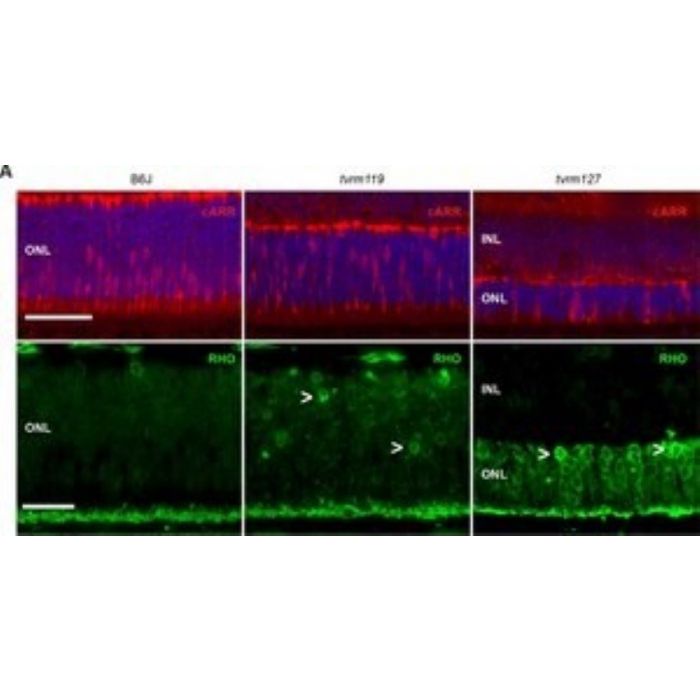
| Background Information | Arrestin-C (UniProt: Q9EQP6; also known as Cone arrestin, cArr, Retinal cone arrestin-3) is encoded by the Arr3 gene (Gene ID: 170735) in murine species. Arrestins are a superfamily of multi-functional proteins that that regulate signaling and trafficking of the majority of G-protein-coupled receptors (GPCRs), as well as sub-cellular localization and activity of many other signaling proteins. Arrestin-C, a disulfide-linked, homodimeric protein, is predominantly found in Inner and outer segments, and the inner plexiform regions of the retina. It is expressed in cone photoreceptors and pinealocytes and may contribute to the shut-off mechanisms associated with high acuity color vision. Arrestin-C is an elongated two-domain molecule with overall fold and key inter-domain interactions that hold the free protein in the basal conformation similar to the other subtypes. Several structural elements are reported to contribute to arrestin binding. The C-terminal acidic region serves a regulatory role in controlling arrestin binding selectivity toward the phosphorylated and activated form of a receptor. The basic N-terminal domain directly participates in receptor interaction and serves a regulatory role via intramolecular interaction with the C-terminal acidic region. Also, two centrally localized domains are directly involved in determining receptor binding specificity and selectivity. Mutations in ARR3 gene in humans have been linked to X-lined myopia 26 that is characterized by typical tigroid fundus changes commonly seen in early onset high myopia. (Ref.: Gurevich, VV., et al. (2018). Protein Cell. 9; 986-1003; Xiao, X., et al. (2016). Mol. Vis. 22; 1257-1266). |
| Product Information | |
|---|---|
| Format | Affinity Purified |
| Presentation | Purified rabbit polyclonal antibody in buffer containing 0.02 M phosphate buffer, pH 7.6, 0.25 M NaCl, and 0.1% sodium azide. |
| Quality Level | MQ100 |
| Applications | |
|---|---|
| Application | Anti-Cone Arrestin, Cat. No. AB15282, is a rabbit polyclonal antibody that detects Arrestin-C and is tested for use in Immunohistochemistry (Paraffin) and Western Blotting. |
| Key Applications |
|
| Application Notes | Tested Applications Immunohistochemistry (Paraffin) Analysis: A 1:500 dilution from a representative lot detected Arrestin-C in Rat retina, Mouse retina and Mouse brain tissue sections. Note: Actual optimal working dilutions must be determined by end user as specimens, and experimental conditions may vary with the end user. |
| Biological Information | |
|---|---|
| Immunogen | KLH-conjugated linear peptide corresponding to 12 amino acids from the C-terminal region of mouse Cone Arrestin. |
| Epitope | C-terminus |
| Concentration | Please refer to lot specific datasheet. |
| Host | Rabbit |
| Specificity | This rabbit polyclonal antibody detects Cone Arrestin. It targets an epitope within 12 amino acids from the C-terminal region. |
| Isotype | IgG |
| Species Reactivity |
|
| Species Reactivity Note | Mouse, Rat |
| Antibody Type | Polyclonal Antibody |
| Entrez Gene Number |
|
| Gene Symbol |
|
| Purification Method | Affinity Purified |
| UniProt Number |
|
| Molecular Weight | ~46 kDa observed; 41.92 kDa calculated. Uncharacterized bands may be observed in some lysate(s). |
| Product Usage Statements | |
|---|---|
| Quality Assurance | Evaluated by Western Blotting in Mouse retina tissue lysate. Western Blotting Analysis: A 1:500 dilution of this antibody detected Cone Arrestin in Mouse retina tissue lysate. |
| Usage Statement |
|
| Storage and Shipping Information | |
|---|---|
| Storage Conditions | Recommended storage: +2°C to +8°C. |
| Packaging Information | |
|---|---|
| Material Size | 100 µg |