

Special Offers
100% Performance Guaranteed
Key Specifications Table
| Species Reactivity | Key Applications | Host | Format | Antibody Type |
|---|---|---|---|---|
| H, R, M, Rhesus Macaque | IH(P), ABA, IF, EM | M | Purified | Monoclonal Antibody |
| Description | |
|---|---|
| Catalogue Number | MAB5456-C |
| Description | Anti-Gonadotropin-Releasing Hormone Antibody, NT, clone HU11B (Ascites Free) |
| Alternate Names |
|
| Background Information | Gonadotropin-releasing hormone I (also known as GnRH, GnRH-I, Gonadoliberin-1, Gonadoliberin I, LHRH, LH-RH I, Luteinizing hormone-releasing hormone I) is encoded by the Gnrh1 (also known as Gnrh, Lhrh1, Lnrh) gene (Gene ID 14714) in murine species. GnRH/LHRH is initially produced as a 90-amino acid progonadoliberin-1 precursor protein (UniProt P13562), removal of the signal peptide (a.a. 1-21) yields progonadoliberin-1 (a.a. 22-90), which is further processed by a proteolytic cleavage to release the GnRH/LHRH decapeptide (a.a. 22-31) and the 56-a.a. prolactin release-inhibiting factor 1 (a.a. 35-90). The LHRH decapeptide functions as the primary endocrine link between the brain and the reproductive system. It plays a primary neuroendocrine role in orchestrating the developmental processes associated with sexual development in immature mammals and also in maintaining normal reproductive function in adults. LHRH-producing neurons are located mainly in the lateral anterior hypothalamus, septum, and medial preoptic area, from where they extend axons towards the median eminence and relese the hormone into the hypothalamo-hypophyseal portal blood vessels in an episodic or pulsatile manner. |
| Product Information | |
|---|---|
| Format | Purified |
| Presentation | Purified mouse monoclonal IgG1κ antibody in buffer containing 0.1 M Tris-Glycine (pH 7.4), 150 mM NaCl with 0.05% sodium azide. |
| Quality Level | MQ100 |
| Applications | |
|---|---|
| Application | Anti-Gonadotropin-Releasing Hormone Antibody, NT, clone HU11B (Ascites Free) is an antibody against Gonadotropin-Releasing Hormone for use in Immunohistochemistry (Paraffin), Affinity Binding Assay, Immunofluorescence, Electron Microscopy. |
| Key Applications |
|
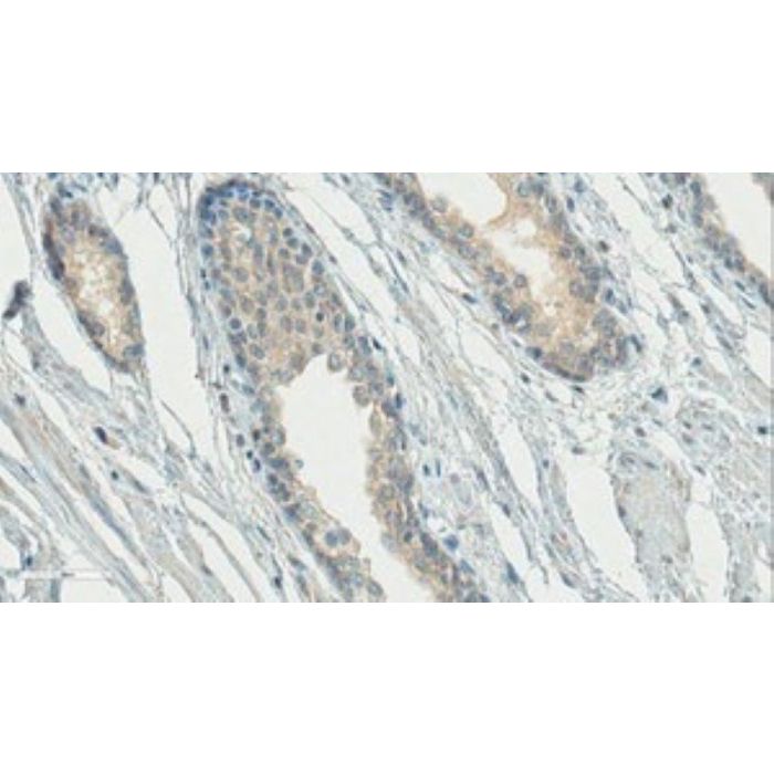
| Application Notes | Immunohistochemistry Analysis: A 1:50 dilution from a representative lot detected Gonadotropin-Releasing Hormone in human prostate cancer tissue. Affinity Binding Assay: A representative lot captured C-terminally blocked GnRH-I/LHRH (LHRH-NH2), but not LHRH with a free C-terminal end (LHRH-COOH). Clone HU11B exhibited reduced affinity toward N-terminally truncated LHRH-NH2 and failed to recognize pro-LHRH, Atrial Naturetic Factor/ANF, Growth hormone-releasing hormone/GHRH, Oxytocin, Somatostatin, Thyrotropin-releasing hormone/TRH, and Vasoactive intestinal peptide/VIP (Urbanski, H.F., et al. (1991). Biol. Reprod. 44(4):681-686). Immunofluorescence Analysis: A representative lot detected GnRH neurons in brain sections from female rhesus monkeys by fluorescent immunohistochemistry (Naugle, M.M., and Gore, A.C. (2014). Neuroendocrinology 100:334-346). Immunofluorescence Analysis: A representative lot detected pregnancy stage-dependent GnRH-I immunoreactivity in the luminal epithelium of rat oviduct by fluorescent immunohistochemistry using paraformaldehyde-fixed cryosections (Sengupta, A., et al. (2007). J. Histochem. Cytochem. 55(5):525-534). Immunofluorescence Analysis: A representative lot detected GnRH-I immunoreactivity in frozen mouse hypothalamic sections by fluorescent immunohistochemistry (Olcese, J., et al. (2003). Neuroreport. 14(4):613-618). Electron Microscopy: A representative lot detected GnRH-I immunoreactivity associated with the luminal epithelium secretory vesicles of rat oviduct by EM using paraformaldehyde-fixed LR wihite sections from rats at day 16 of pregnancy (Sengupta, A., et al. (2007). J. Histochem. Cytochem. 55(5):525-534). Immunohistochemistry Analysis: A representative lot detected GnRH-I immunoreactivity throughout the hypothalamus in paraformalin-fixed, paraffin-embedded South American plains vizcacha (Lagostomus maximus) brain sections (Dorfman, V.B., et al. (2011). J. Mol. Histol. 42(4):311-321). Immunohistochemistry Analysis: A representative lot detected GnRH-I-positie neurons in formalin-fixed, paraffin-embedded mouse brain sections (Bergman, J.E., et al. (2010). Eur. J. Hum. Genet. 18(2):171-177). Immunohistochemistry Analysis: A representative lot detected GnRH-I immunoreactivity in paraformaldehyde-fixed rat brain vibratome sections (Urbanski, H.F., et al. (1991). Biol. Reprod. 44(4):681-686). |
| Biological Information | |
|---|---|
| Immunogen | Mouse GnRH (QHWSYGLRPG) conjugated to bovine thyroglobulin. |
| Epitope | N-terminus. |
| Clone | HU11B |
| Concentration | Please refer to lot specific datasheet. |
| Host | Mouse |
| Specificity | Clone HU11B reacts with C-terminally blocked GnRH-I/LHRH (LHRH-NH2), but not LHRH with a free C-terminal end (LHRH-COOH). Clone HU11B exhibits reduced affinity toward N-terminally truncated LHRH-NH2 and failed to recognize pro-LHRH, Atrial Naturetic Factor/ANF, Growth hormone-releasing hormone/GHRH, Oxytocin, Somatostatin, Thyrotropin-releasing hormone/TRH, and Vasoactive intestinal peptide/VIP (Urbanski, H.F., et al. (1991). Biol. Reprod. 44(4):681-686). |
| Isotype | IgG1κ |
| Species Reactivity |
|
| Antibody Type | Monoclonal Antibody |
| Entrez Gene Number |
|
| Gene Symbol |
|
| Purification Method | Protein G Purified |
| UniProt Number |
|
| Molecular Weight | 10 kDa calculated |
| Product Usage Statements | |
|---|---|
| Quality Assurance | Evaluated by Immunohistochemistry in human prostate tissue. Immunohistochemistry Analysis: A 1:50 dilution of this antibody detected Gonadotropin-Releasing Hormone in human prostate tissue. |
| Usage Statement |
|
| Storage and Shipping Information | |
|---|---|
| Storage Conditions | Stable for 1 year at 2-8°C from date of receipt. |
| Packaging Information | |
|---|---|
| Material Size | 100 μg |