

Special Offers
100% Performance Guaranteed
Key Specifications Table
| Species Reactivity | Key Applications | Host | Format | Antibody Type |
|---|---|---|---|---|
| Vrt | ICC, WB | Ch | Purified | Polyclonal Antibody |
| Description | |
|---|---|
| Catalogue Number | 06-896 |
| Brand Family | Upstate |
| Trade Name |
|
| Description | Anti-GFP (Green Fluorescent Protein) Antibody |
| Alternate Names |
|
| Background Information | Green fluorescent protein (GFP) is a 29 kDa fluorescent protein isolated from the jellyfish Aequorea victoria. Upon exposure to blue light (488 nm), GFP strongly fluoresces (Emission max: 507 nm). GFP can be fused to proteins without significantly interfering with their assembly and function, making GFP a powerful tool for monitoring gene expression and protein localization in living cells. |
| Product Information | |
|---|---|
| Format | Purified |
| Control |
|
| Presentation | Purified chicken polyclonal IgY from egg yolk in buffer containing 70% storage buffer (0.1M Tris-glycine, pH 7.4, 0.15M NaCl, 0.05% sodium azide) and 30% glycerol. Store at -20°C. |
| Quality Level | MQ100 |
| Applications | |
|---|---|
| Application | Anti-GFP (Green Fluorescent Protein) Antibody detects level of GFP (Green Fluorescent Protein) & has been published & validated for use in IC & WB. |
| Key Applications |
|
| Application Notes | Immunocytochemistry: This antibody was reported to show positive immunostaining in cells transfected with an expression vector encoding Green Fluorescent Protein. |
| Biological Information | |
|---|---|
| Immunogen | His-tagged green fluorescent protein of Aequorea victoria. |
| Concentration | Please refer to the Certificate of Analysis for the lot-specific concentration. |
| Host | Chicken |
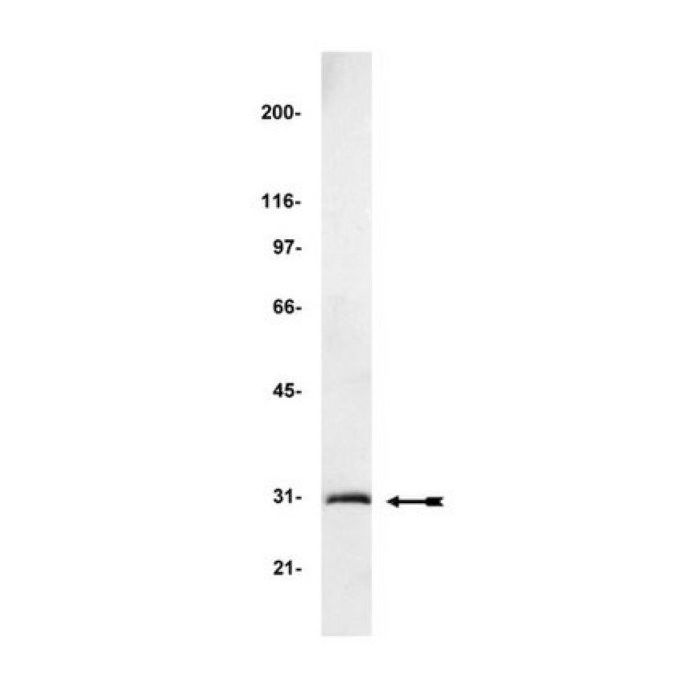
| Specificity | Recognizes green fluorescent protein (GFP), Mr 30 kDa, and GFP-fusion proteins. |
| Isotype | IgY |
| Species Reactivity |
|
| Antibody Type | Polyclonal Antibody |
| Gene Symbol |
|
| Purification Method | Ammonium Sulfate Precipitation and PEG |
| UniProt Number |
|
| UniProt Summary | FUNCTION: Energy-transfer acceptor. Its role is to transduce the blue chemiluminescence of the protein aequorin into green fluorescent light by energy transfer. Fluoresces in vivo upon receiving energy from the Ca(2+)-activated photoprotein aequorin. BIOPHYSICOCHEMICAL PROPERTIES: Absorption: Abs(max)=395 nm; Note=Exhibits a smaller absorbance peak at 470 nm. The fluorescence emission spectrum peaks at 509 nm with a shoulder at 540 nm; SUBUNIT: Monomer. TISSUE SPECIFICITY: Photocytes. PTM: Contains a chromophore consisting of modified amino acid residues. The chromophore is formed by autocatalytic backbone condensation between Ser-65 and Gly-67, and oxidation of Tyr-66 to didehydrotyrosine. Maturation of the chromophore requires nothing other than molecular oxygen. BIOTECHNOLOGY: Green fluorescent protein has been engineered to produce a vast number of variously colored mutants, fusion proteins, and biosensors. Fluorescent proteins and its mutated allelic forms, blue, cyan and yellow have become a useful and ubiquitous tool for making chimeric proteins, where they function as a fluorescent protein tag. Typically they tolerate N- and C-terminal fusion to a broad variety of proteins. They have been expressed in most known cell types and are used as a noninvasive fluorescent marker in living cells and organisms. They enable a wide range of applications where they have functioned as a cell lineage tracer, reporter of gene expression, or as a measure of protein-protein interactions. BIOTECHNOLOGY: Can also be used as a molecular thermometer, allowing accurate temperature measurements in fluids. The measurement process relies on the detection of the blinking of GFP using fluorescence correlation spectroscopy. SIMILARITY: Belongs to the GFP family. |
| Molecular Weight | 30 kDa |
| Product Usage Statements | |
|---|---|
| Quality Assurance | Routinely evaluated by Western Blot on rat brain lysate containing purified GFP (Catalog #14-392) or a GFP-fusion protein from a transfected E. coli cell lysate. Western Blot Analysis: 0.5-2 μg/mL of this lot detected Green Fluorescent Protein from 50 μg of rat brain lysate containing 25 ng of purified Green Fluorescent Protein (Catalog # 14-392). A previous lot of this antibody also detected a GFP-fusion protein from a transfected E. coli cell lysate. |
| Usage Statement |
|
| Storage and Shipping Information | |
|---|---|
| Storage Conditions | Stable for 1 year at -20°C from date of receipt. Handling Recommendations: Upon receipt, and prior to removing the cap, centrifuge the vial and gently mix the solution. Aliquot into microcentrifuge tubes and store at -20°C. Avoid repeated freeze/thaw cycles, which may damage IgG and affect product performance. Note: Variability in freezer temperatures below -20°C may cause glycerolcontaining solutions to become frozen during storage. |
| Packaging Information | |
|---|---|
| Material Size | 200 µg |