We use cookies to make your experience better. To comply with the new e-Privacy directive, we need to ask for your consent to set the cookies. Learn more.
Share this :
Print
Cell Signaling Pdap1 Antibody
Cell Signaling Pdap1 Antibody - CSIG (Additional S&H or Hazmat Fees May Apply)
List Price
$302.00
Your Price
$302.00
HOW MUCH YOU SAVE:
0.00 %


| NETA PART: | CSIG-4300S |
| MFG.PART: | 4300S |
| UNSPSC: | 12352203 |
| Manufacturer: | Cell Signaling |
| Size | 100 µl |
| Reactivity | H M R Mk |
| Sensitivity | Endogenous |
| Molecular Weight (kDa) | 25 |
| Source/Isotype | Rabbit |
| Application/Dilution | {Western Blotting: 1:1000, Immunoprecipitation: 1:50} |
| Storage | Supplied in 10 mM sodium HEPES (pH 7.5), 150 mM NaCl, 100 µg/ml BSA and 50% glycerol. Store at –20°C. Do not aliquot the antibody. |
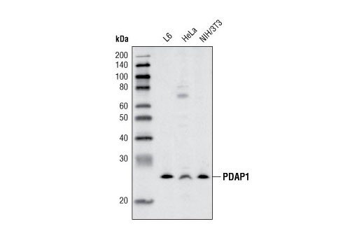
| Specificity/Sensitivity | PDAP1 Antibody detects endogenous levels of total PDAP1 protein. Nonspecific bands are seen above 60 KDa. |
| Species Reactivity | Human, Mouse, Rat, Monkey |
| Source/Purification | Polyclonal antibodies are produced by immunizing animals with a synthetic peptide corresponding to residues near the center of human PDAP1 protein. Antibodies were purified by protein A and peptide affinity chromatography. |
| Background | Platelet derived growth factor (PDGF) proteins function as dimeric isoforms (i.e., PDGF AA, PDGF AB, PDGF BB, PDGF CC and PDGF DD) that bind receptor tyrosine kinases and activate cytoplasmic SH2 domain-containing proteins to control multiple signaling pathways that regulate angiogenesis, cell growth, actin reorganization, migration and differentiation (1). PDGFA-associated protein 1 (PDAP1) was originally identified as a novel, PDGF-associated protein found in a rat retinal tumor cell line (2). While copurified with PDGFA, PDAP1 interacts with PDGFB at a slightly higher affinity than with PDGFA (2). Although the exact function of PDAP1 is unclear, it has been shown to both increase PDGFA-induced incorporation of [3H]thymidine in Swiss 3T3 cells and decrease PDGFB growth factor activity (2). Ubiquitously expressed PDAP1 is highly conserved among species (2,3) and is phosphorylated in vitro by several kinases, including PKC, PKA, CKI and CKII. Among this group, CKII seems to be the major kinase that phosphorylates PDAP1 in intact cells (3). |
| SKU | CSIG-4300S |
|---|---|
| Supplier Part Number | 4300S |
| UM | EA |
| UNSPSC | 12352203 |
| Manufacturer | Cell Signaling |
| MSDS URL | https://www.cellsignal.com/contents/technical/safety-data-sheet-(sds)/resources-safety-data-sheets |
| Temperature | -20C |
| ProductLine | CSIG |
| Qty | 1 |
| MinOrderQty | 1 |
| Weight | 7.00 |
| Lead Time | 5 Business Days |
| Hazardous | N |