We use cookies to make your experience better. To comply with the new e-Privacy directive, we need to ask for your consent to set the cookies. Learn more.
Share this :
Print
Cell Signaling Ddb-1 Antibody
Cell Signaling Ddb-1 Antibody - CSIG (Additional S&H or Hazmat Fees May Apply)
List Price
$358.00
Your Price
$358.00
HOW MUCH YOU SAVE:
0.00 %


| NETA PART: | CSIG-5428S |
| MFG.PART: | 5428S |
| UNSPSC: | 12352203 |
| Manufacturer: | Cell Signaling |
| Size | 100 µl |
| Reactivity | H M R Mk |
| Sensitivity | Endogenous |
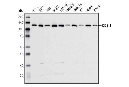
| Molecular Weight (kDa) | 127 |
| Source/Isotype | Rabbit |
| Application/Dilution | {Western Blotting: 1:1000} |
| Storage | Supplied in 10 mM sodium HEPES (pH 7.5), 150 mM NaCl, 100 µg/ml BSA and 50% glycerol. Store at –20°C. Do not aliquot the antibody. |
| Specificity/Sensitivity | DDB-1 Antibody detects endogenous levels of total DDB-1 protein. |
| Species Reactivity | Human, Mouse, Rat, Monkey |
| Source/Purification | Polyclonal antibodies are produced by immunizing animals with a synthetic peptide corresponding to residues near the carboxy terminus of human DDB-1 protein. Antibodies are purified by protein A and peptide affinity chromatography. |
| Background | Damaged DNA-Binding Protein (DDB) consists of a 127 kDa subunit (DDB-1) and a 48 kDa subunit (DDB-2) that contribute to the formation of the UV-damaged DNA-binding protein complex (UV-DDB) (1-3). In conjunction with CUL4A and ROC-1, the UV-DDB complex forms an E3 ubiquitin ligase that recognizes a broad spectrum of DNA lesions such as cyclobutane pyrimidine dimers, 6-4 photoproducts, apurinic sites and short mismatches. The complex polyubiquitinates components of the nucleotide excision repair pathway (4-6). Loss of DDB activity has been identified in a subset of xeroderma pigmentosum complementation group E (XP-E) patients and has been linked to the deficient repair of cyclobutane pyrimidine dimers in cells derived from these patients (7-10).DDB-1 is a relatively abundant protein that is vital for normal cell function and is evolutionarily conserved in mammals, insects, worms and plants. Unlike DDB-2, lesions in DDB-1 have yet to be indentified in XP-E patients. In association with ROC-1 and CUL4A, DDB-1 functions to recruit substrate-specific targeting subunits, generally known as DCAFs or CDWs, to CUL4-RING E3 ubiquitin-protein ligase complexes (11,12). Ubiquitination of histone H2A, histone H3 and histone H4 at sites of UV-induced DNA damage by the DDB1-DDB2-CUL4A-ROC1 E3 ubiquitin-protein ligase complex may facilitate their removal from the nucleosome in order to promote DNA repair (13-15). DDB-1, in association with other CUL4-based E3 ligase complexes, has also been found to be a regulator of mTOR signaling (16,17). |
| SKU | CSIG-5428S |
|---|---|
| Supplier Part Number | 5428S |
| UM | EA |
| UNSPSC | 12352203 |
| Manufacturer | Cell Signaling |
| MSDS URL | https://www.cellsignal.com/contents/technical/safety-data-sheet-(sds)/resources-safety-data-sheets |
| Temperature | -20C |
| ProductLine | CSIG |
| Qty | 1 |
| MinOrderQty | 1 |
| Weight | 7.00 |
| Lead Time | 5 Business Days |