

Special Offers
100% Performance Guaranteed
Key Specifications Table
| Species Reactivity | Key Applications | Host | Format | Antibody Type |
|---|---|---|---|---|
| M, H | WB, IH(P) | M | Purified | Monoclonal Antibody |
| Description | |
|---|---|
| Catalogue Number | MABS471 |
| Description | Anti-CYP26C1 Antibody, clone T6P1C7*E7 |
| Alternate Names |
|
| Background Information | Cytochrome P450 26C1 (UniProt Q6V0L0; also known as CYP26C1, Cytochrome P450, family 26, subfamily C, polypeptide 1) is encoded by the CYP26C1 (also known as FFDD4) gene (Gene ID 340665) in human. Cytochromes P450 (CYP) proteins are primarily membrane-associated oxidases located either in the inner membrane of mitochondria or in the endoplasmic reticulum where they function as the terminal enzymes in electron transfer chains. In addition to processing endogenous substrates, CYPs also function to metabolize exogenous drugs and potentially toxic chemicals. The human CYP superfamily consisits of 57 genes and more than 59 pseudogenes divided into 18 families and 43 subfamilies. The CYP26 subfamily of enzymes (CYP26A1, CYP26B1/CYP26A2, CYP26C1) are retinoic acid hydroxylases responsible for the inactivation of all-trans-retinoic acid (atRA) to hydroxylated forms, such as 4-oxo-, 4-OH-, and 18-OH-atRA, with 4-oxo-RA being the most common metabolite. All-trans-RA represents the most active form of RA and plays a crucial role in the development of multiple organs via its gene regulatory function. 4-oxo-9-cis-retinoic acid (9-cis-RA) and 4-oxo-13-cis-retinoic acid (13-cis-RA) are two atRA stereo-isomers that also play an important role in RA signalling. Immunohistochemistry staining reveals moderate to strong expression of CYP26A1 and CYP26B1 in colon cancer tissues when compared with normal colonic epithelium, while CYP26C1 was not expressed in either type of colon tissue samples. CYP26C1 gene mutations are linked to focal facial dermal dysplasia 4 (FFDD4), a group of developmental defects characterized by bitemporal or preauricular skin lesions resembling aplasia cutis congenita. |
| Product Information | |
|---|---|
| Format | Purified |
| Presentation | Purified mouse monoclonal IgG1 in buffer containing 0.1 M Tris-Glycine (pH 7.4) 150 mM NaCl with 0.05% sodium azide. |
| Quality Level | MQ100 |
| Applications | |
|---|---|
| Application | This mouse monoclonal Anti-CYP26C1 Antibody, clone T6P1C7*E7, Cat. No. MABS471 detects levels of CYP26C1, and has been published and validated for use in ELISA, Immunohistochemistry (Paraffin), and Western Blotting. |
| Key Applications |
|
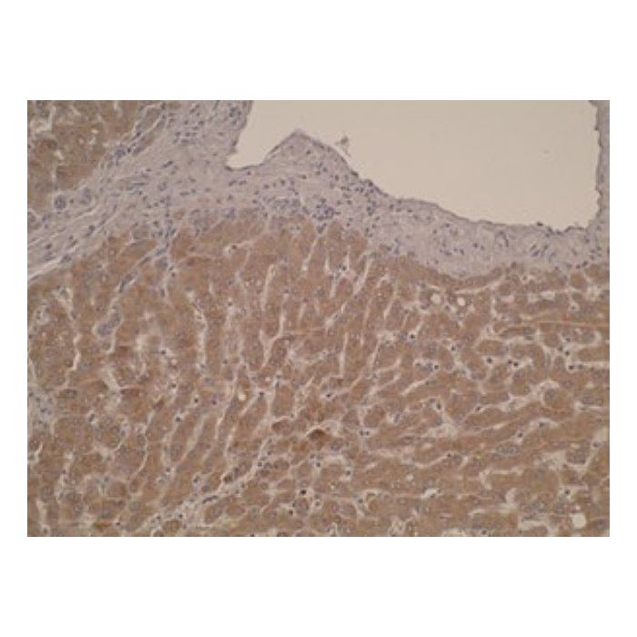
| Application Notes | Immunohistochemistry Analysis: Clone T6P1C7*E7 hybridoma culture supernatant detected cytoplasmic CYP51A1 immunoreactivity in formalin-fixed, paraffin-embedded human liver tissue sections. ELISA Analysis: Clone T6P1C7*E7 hybridoma culture supernatant deteced ovalbumin-conjugated immunogen peptide by ELISA (Brown, G.T., et al. (2014). PLoS One. 9(3):e90776). Western Blotting Analysis: Clone T6P1C7*E7 hybridoma culture supernatant deteced the expression of exogenously transfected CYP26C1 in lysates from transfected, but not mock-transfected, human embryonic kidney cells (Brown, G.T., et al. (2014). PLoS One. 9(3):e90776). |
| Biological Information | |
|---|---|
| Immunogen | Ovalbumin-conjugated linear peptide corresponging to a C-terminal region sequence of human/mouse/rat CYP26C1. |
| Clone | T6P1C7*E7 |
| Concentration | Please refer to lot specific datasheet. |
| Host | Mouse |
| Specificity | Clone T6P1C7*E7 was raised against a CYP26C1 C-terminal sequence 100% conserved among human, mouse, and rat species. |
| Isotype | IgG1κ |
| Species Reactivity |
|
| Species Reactivity Note | Mouse, Human. Predicted to react with Rat based on 100% sequence homology. |
| Antibody Type | Monoclonal Antibody |
| Entrez Gene Number |
|
| Gene Symbol |
|
| Purification Method | Protein G purified. |
| UniProt Number |
|
| Molecular Weight | ~57 kDa observed. 57.11/57.03/57.30 kDa (human/mouse/rat) calculated. Uncharacterized bands may be observed in some lysate(s). |
| Product Usage Statements | |
|---|---|
| Quality Assurance | Evaluated by Western Blotting in human liver microsome lysate. Western Blotting Analysis: 10 µg/mL of this antibody detected CYP26C1 in 50 µg of human liver microsome lysate. |
| Usage Statement |
|
| Storage and Shipping Information | |
|---|---|
| Storage Conditions | Stable for 1 year at 2-8°C from date of receipt. |
| Packaging Information | |
|---|---|
| Material Size | 100 μg |