

Key Specifications Table
| Species Reactivity | Key Applications | Host | Format | Antibody Type |
|---|---|---|---|---|
| M, H | ELISA, FC, IH(P), WB | M | Purified | Monoclonal Antibody |
| Description | |
|---|---|
| Catalogue Number | MABT858-25UG |
| Description | Anti-prelamin A Antibody, clone PL-1C7 |
| Alternate Names |
|
| Background Information | Prelamin-A/C (UniProt: P02545; also known as 70 kDa lamin, Renal carcinoma antigen NY-REN-32) is encoded by the LMNA (also known as LMN1) gene (Gene ID: 4000) in human. Lamins are components of the nuclear lamina that provide a framework for the nuclear envelope and may also interact with chromatin. Lamin A and C are present in equal amounts in the lamina of mammals. Plays an important role in nuclear assembly, chromatin organization, nuclear membrane and telomere dynamics. Lamin A is initially synthesized as prelamin A that undergoes several modifications in the carboxyl terminal region that allow incorporation of prelamin A into the nuclear envelope and its subsequent processing into the mature lamin A. Cleavage of 15 residues (aa 647-662) by ZMPSTE24/FACE1 generates the final protein product. Unlike mature lamin A, prelamin A accumulates as discrete and localized foci at the nuclear periphery. Prelamin-A/C can accelerate smooth muscle cell senescence. It can act to disrupt mitosis and induce DNA damage in vascular smooth muscle cells (VSMCs), leading to mitotic failure, genomic instability, and premature senescence. Mutations in LMNA gene are known to cause Emery-Dreifuss muscular dystrophy that is characterized by weakness and atrophy of muscle without involvement of the nervous system. Some mutations have also been linked to familial type of lipodystrophy characterized by the loss of subcutaneous adipose tissue in the lower parts of the body. (Ref.: Casasola, A., et al. (2016). Nucleus 7(1); 84-102). |
| Product Information | |
|---|---|
| Format | Purified |
| Presentation | Purified mouse monoclonal antibody IgG2b in buffer containing 0.1 M Tris-Glycine (pH 7.4), 150 mM NaCl with 0.05% sodium azide. |
| Applications | |
|---|---|
| Application | Anti-prelamin A, clone PL-1C7, Cat. No. MABT858, is a mouse monoclonal antibody that detects Prelamin A and has been tested for use in ELISA, Flow Cytometry, Immunocytochemistry, and Western Blotting. |
| Key Applications |
|
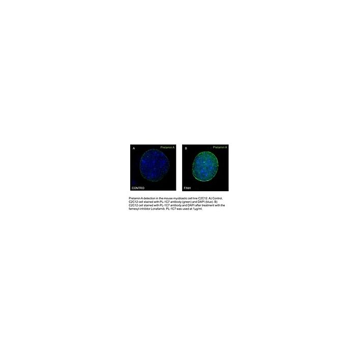
| Application Notes | Western Blotting Analysis: 2 µg/mL from a representative lot detected prelamin A in U20S cells treated with farnesyl transferase inhibitor, Lonafarnib (3.2 uM for 24 h) . Flow Cytometry Analysis: A representative lot detected prelamin A in Flow Cytometry applications (Casasola, A., et. al. (2016). Nucleus. 7(1):84-102). Western Blotting Analysis: A representative lot detected prelamin A in Western Blotting applications (Casasola, A., et. al. (2016). Nucleus. 7(1):84-102). Immunocytochemistry Analysis: 1 µg/mL from a representative lot detected prelamin A in C2C12 cells with Farnesyl transferase inhibitor Lonafarnib (Courtesy of Fred Hutchinson Cancer Research Center, Seattle, Washington USA). ELISA Analysis: A representative lot detected prelamin A in ELISA applications (Casasola, A., et. al. (2016). Nucleus. 7(1):84-102). Immunocytochemistry Analysis: A representative lot detected prelamin A in Immunocytochemistry applications (Casasola, A., et. al. (2016). Nucleus. 7(1):84-102). |
| Biological Information | |
|---|---|
| Immunogen | A lnear peptide corresponding to 12 amino acids from the C-terminal region of human Prelamin A. |
| Clone | PL-1C7 |
| Concentration | Please refer to lot specific datasheet. |
| Host | Mouse |
| Specificity | Clone PL-1C7 detects Prelamin A in human and murine cells. It specifically recognizes prelamin A at the intact ZMPSTE24 cleavage site. |
| Isotype | IgG2b |
| Species Reactivity |
|
| Species Reactivity Note | Human, Mouse. |
| Antibody Type | Monoclonal Antibody |
| Gene Symbol |
|
| Purification Method | Protein G purified |
| UniProt Number |
|
| Molecular Weight | ~74 kDa observed; 74.14 kDa calculated. Uncharacterized bands may be observed in some lysate(s). |
| Product Usage Statements | |
|---|---|
| Quality Assurance | Evaluated by Western Blotting in C2C12 cell lysate. Western Blotting Analysis: 2 µg/mL of this antibody detected prelamin A in C2C12 cell lysate. |
| Usage Statement |
|
| Storage and Shipping Information | |
|---|---|
| Storage Conditions | Stable for 1 year at 2-8°C from date of receipt. |
| Packaging Information | |
|---|---|
| Material Size | 25 μg |