We use cookies to make your experience better. To comply with the new e-Privacy directive, we need to ask for your consent to set the cookies. Learn more.
Share this :
Print
Cell Signaling Pd-L1 (E1l3n ) Xp Rabbit mAb
Cell Signaling Pd-L1 (E1l3n ) Xp Rabbit mAb - CSIG (Additional S&H or Hazmat Fees May Apply)
List Price
$173.00
Your Price
$173.00
HOW MUCH YOU SAVE:
0.00 %


| NETA PART: | CSIG-13684T |
| MFG.PART: | 13684T |
| UNSPSC: | 12352203 |
| Manufacturer: | Cell Signaling |
| Size | 20 µl |
| Reactivity | H |
| Sensitivity | Endogenous |
| Molecular Weight (kDa) | 40-50 |
| Source/Isotype | Rabbit IgG |
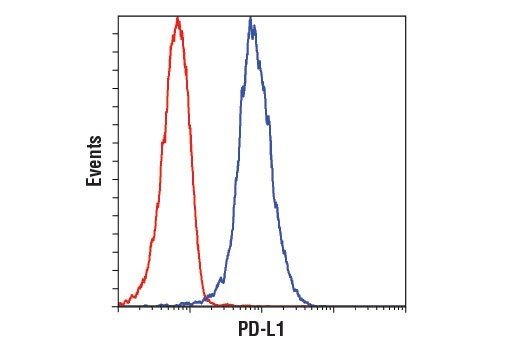
| Application/Dilution | {Western Blotting: 1:1000, Immunoprecipitation: 1:50, IHC Leica Bond: 1:200 - 1:800, Immunohistochemistry (Paraffin): 1:100 - 1:400, Flow Cytometry (Fixed/Permeabilized): 1:200 - 1:800} |
| Storage | Supplied in 10 mM sodium HEPES (pH 7.5), 150 mM NaCl, 100 µg/ml BSA, 50% glycerol and less than 0.02% sodium azide. Store at –20°C. Do not aliquot the antibody.For a carrier free (BSA and azide free) version of this product see product #85164. |
| Specificity/Sensitivity | PD-L1 (E1L3N®) XP® Rabbit mAb recognizes endogenous levels of total PD-L1 protein. |
| Species Reactivity | Human |
| Source/Purification | Monoclonal antibody is produced by immunizing animals with a synthetic peptide corresponding to residues near the carboxy terminus of human PD-L1 protein. |
| Background | Programmed cell death 1 ligand 1 (PD-L1, B7-H1, CD274) is a member of the B7 family of cell surface ligands that regulate T cell activation and immune responses. The PD-L1 ligand binds the PD-1 transmembrane receptor and inhibits T cell activation. PD-L1 was discovered following a search for novel B7 protein homologs and was later shown to be expressed by antigen presenting cells, activated T cells, and tissues including placenta, heart, and lung (1-3). Similar in structure to related B7 family members, PD-L1 protein contains extracellular IgV and IgC domains and a short, cytoplasmic region. Research studies demonstrate that PD-L1 is expressed in several tumor types, including melanoma, ovary, colon, lung, breast, and renal cell carcinomas (4-6). Expression of PD-L1 in cancer is associated with tumor-infiltrating lymphocytes, which mediate PD-L1 expression through the release of interferon gamma (7). Additional research links PD-L1 expression to cancers associated with viral infections (8,9). |
| SKU | CSIG-13684T |
|---|---|
| Supplier Part Number | 13684T |
| UM | EA |
| UNSPSC | 12352203 |
| Manufacturer | Cell Signaling |
| MSDS URL | https://www.cellsignal.com/contents/technical/safety-data-sheet-(sds)/resources-safety-data-sheets |
| Temperature | -20C |
| ProductLine | CSIG |
| Qty | 1 |
| MinOrderQty | 1 |
| Weight | 7.00 |
| Lead Time | 5 Business Days |